If you are looking to source blood products for your life science research, choosing the right anticoagulant can be daunting. In this blog, we will provide an overview of the most used anticoagulants, their applications, advantages and disadvantages.
So, let’s start with the basics…
How do anticoagulants work?
Anticoagulation occurs in the two following ways:
- Binding calcium ions (e.g. EDTA, citrate)
- Inhibiting thrombin activity (e.g. heparin)
The three most used anticoagulants are:
- Ethylenediaminetetraacetic acid (EDTA)
- Heparin
- Citrate
How can anticoagulants interfere with analysis?
The coagulation process can change the concentrations of various constituents in extracellular fluid, pushing them beyond their maximum allowable limits. To obtain clinically relevant results, certain constituents should only be measured in plasma, such as ammonia, serotonin and neuron-specific enolase.
Therefore, adding anticoagulants can interfere with certain analytical methods or alter the concentrations of the constituents to be measured, such as:
- Contamination with cations: NH4+, Li+, Na+, K+
- Assay interference: Caused by metals complexing with EDTA and citrate, for example:
- Inhibition of alkaline phosphatase activity by zinc binding
- Inhibition of metalloproteinases
- Inhibition of metal-dependent cell activation in function tests
- Binding of ionised calcium to heparin
- Interference by fibrinogen: In heterogeneous immunoassays
- Inhibition of metabolic or catalytic reactions by heparin: For example, Taq polymerase in the polymerase chain reaction (PCR)
- Interference in ion distribution: Between the intracellular and extracellular spaces, such as Cl-, NH4+ by EDTA and citrate
Can anticoagulants affect PCR assays?
Yes. Anticoagulants, more specifically those that are heparin-based, can inhibit downstream PCR assays. PCR analysis can be applied to a wide variety of biospecimens, and the following precautions must be taken when using heparinised material where other anticoagulants cannot be used.
- Simple PCR tests: For simple PCR tests which do not require high sensitivity, dilution of the prepared nucleic acids is usually sufficient to overcome the inhibition. If heparinised material must be used and a more sensitive DNA PCR is required, nucleated cells should be isolated first, then washed repeatedly in physiological buffers before further processing
- Highly sensitive RT-PCR methods: For highly sensitive RT-PCR methods, additional measures are necessary to overcome heparin inhibition. Less effective methods include boiling, Sephadex chromatography, pH shifts with subsequent gel filtration, repeated ethanol precipitations, and treatment with protamine sulphate. Although treatment with heparinase restores amplification, this enzymatic purification step is costly. Additionally, RNA may be degraded during enzyme incubation by traces of RNase still present in the sample or by heparinase preparations contaminated with RNase
- Lithium chloride method: Recently, it has been demonstrated that lithium chloride can separate heparin from RNA, thus reversing the inhibition. This method, which reliably restores amplification from heparinised blood samples, is easily incorporated into a routine RNA preparation procedure without additional effort and so would be the preferred method
Anticoagulant selection guide
The following table includes commonly used anticoagulants, appropriate applications, and why:
- If you are using a mobile device, scroll left and right to view all columns
|
Anticoagulant |
Most suitable applications |
Not recommended for |
Advantages |
Disadvantages |
|
K2EDTA / K3EDTA |
|
|
|
|
|
Lithium/sodium heparin |
|
|
|
|
|
Sodium citrate |
|
|
|
|
|
ACD-A |
|
|
|
|
|
CPD |
|
|
|
|
|
Fluoride / oxalate (mixture of potassium oxalate & sodium fluoride) |
|
|
|
|
When to choose serum for your research application: metabolomics studies
For the reasons mentioned earlier, serum (the portion of plasma remaining after blood clotting in the absence of any anticoagulants) is recommended for metabolomics studies to avoid interference from anticoagulants in downstream applications. Serum is widely used for serological diagnosis of infectious diseases using multiple techniques including immunodiffusion, immunoprecipitation, counter immunoelectrophoresis, bacterial agglutination, haemagglutination and agglutination inhibition, particle-enhanced agglutination, complement fixation, indirect immunofluorescence (IFA), enzyme-linked immunoassay (ELISA), radioimmunoassay (RIA), neutralisation of toxins or virus activity, immunoblot (Western blot), etc.
For other tests, including some haemagglutination tests, ELISAs, or immunoblots, either serum or plasma may be used.
If serum is unavailable, both heparin plasma and EDTA plasma approximate the concentrations observed in serum closely.
Considerations for using gel separator tubes in metabolomics
An important distinction needs to be made between conventional blood collection tubes and gel separator tubes for metabolomic analysis. Gel separator tubes are used to accelerate the process of serum or plasma separation and due to the inert gel used, should not change the metabolite composition. However, several studies have highlighted differences in the metabolite fingerprints of samples collected using gel tubes compared to conventional tubes, particularly for amino acids. Hence, the use of gel separator tubes is not recommended.
Meeting your exact research project needs
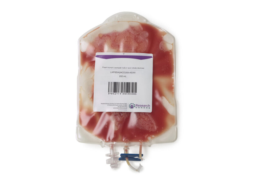
We ensure you get the Right Donor, the Right Sample at the Right Time for your research. We collect from a large diverse pool of donors. Sample types include whole blood, leukopaks, PBMCs, buffy coat, plasma, serum and much more.
All samples are collected under a comprehensive informed consent for commercial and genetic research, and are processed to the highest quality standards in an ISO accredited and HTA (Human Tissue Authority) licenced facility
If you would like to talk to us about your requirements, please e-mail administration@researchdonors.co.uk or call +44 (0) 207 870 3742
References & further reading
- Betsou, F., Gaignaux, A., Ammerlaan, W., Norris, P.J. and Stone, M. (2019). Biospecimen Science of Blood for Peripheral Blood Mononuclear Cell (PBMC) Functional Applications. Current Pathobiology Reports, 7(2), pp.17–27. doi:https://doi.org/10.1007/s40139-019-00192-8.
- Bowen, R.A.R. and Remaley, A.T. (2014). Interferences from blood collection tube components on clinical chemistry assays. Biochemia Medica, 24(1), pp.31–44. doi:https://doi.org/10.11613/bm.2014.006.
- Caliezi, C., Reber, G., Lämmle, B., de Moerloose, P. and Wuillemin, W.A. (2000). Agreement of D-dimer results measured by a rapid ELISA (VIDAS) before and after storage during 24h or transportation of the original whole blood samples. Thrombosis and Haemostasis, [online] 83(1), pp.177–178. Available at: https://pubmed.ncbi.nlm.nih.gov/10669177/ [Accessed 29 May 2024].
- Chowdhury, F.R., Rodman, H. and Bleicher, S. (1971). Glycerol-like contamination of commercial blood sampling tubes. Journal of Lipid Research, [online] 12(1), p.116. Available at: https://pubmed.ncbi.nlm.nih.gov/5542696/#:~:text=Abstract [Accessed 29 May 2024].
- England, J.M., Rowan, R.M., van Assendelft, O.W., Bull, B.S., Coulter, W., Fujimoto, K., Groner, W., Richardson-Jones, A., Klee, G., Koepke, J.A., Lewis, S.M., McLaren, C.E., Shinton, N.K., Tatsumi, N. and Verwilghen, R.L. (1993). Recommendations of the International Council for Standardization in Haematology for Ethylenediaminetetraacetic Acid Anticoagulation of Blood for Blood Cell Counting and Sizing: International Council for Standardization in Haematology: Expert Panel on Cytometry. American Journal of Clinical Pathology, 100(4), pp.371–372. doi:https://doi.org/10.1093/ajcp/100.4.371.
- Ladenson, J.H., L. M.B. Tsai, Michael, J.M., Kessler, G. and J. Heinrich Joist (1974). Serum versus Heparinized Plasma for Eighteen Common Chemistry Tests: Is Serum the Appropriate Specimen? Am J Clinical Pathology, 62(4), pp.545–552. doi:https://doi.org/10.1093/ajcp/62.4.545.
- Leonard, P.J., Persaud, J. and Motwani, R. (1971). The estimation of plasma albumin by BCG RCG binding on the technicon SMA 12/60 analyser and a comparison with the HABA dye binding technique. Clinica Chimica Acta, 35(2), pp.409–412. doi:https://doi.org/10.1016/0009-8981(71)90214-2.
- Li, G., Cabanero, M., Wang, Z., Wang, H., Huang, T., Alexis, H., Eid, I., Muth, G. and Pincus, M.R. (2013). Comparison of glucose determinations on blood samples collected in three types of tubes. Annals of Clinical and Laboratory Science, [online] 43(3), pp.278–284. Available at: https://pubmed.ncbi.nlm.nih.gov/23884222/ [Accessed 29 May 2024].
- Meng, Q.H. and Krahn, J. (2008). Lithium heparinised blood-collection tubes give falsely low albumin results with an automated bromcresol green method in haemodialysis patients. Clinical Chemical Laboratory Medicine, 46(3). doi:https://doi.org/10.1515/cclm.2008.079.
- Narayanan, S. (2000). The Preanalytic Phase. American Journal of Clinical Pathology, 113(3), pp.429–452. doi:https://doi.org/10.1309/c0nm-q7r0-ll2e-b3uy.
- Neumaier, M., Braun, A. and Wagener, C. (1998). Fundamentals of quality assessment of molecular amplification methods in clinical diagnostics. International Federation of Clinical Chemistry Scientific Division Committee on Molecular Biology Techniques. Clinical Chemistry, [online] 44(1), pp.12–26. Available at: https://pubmed.ncbi.nlm.nih.gov/9550553/.
- Numata, Y., Dohi, K., Furukawa, A., Kikuoka, S., Asada, H., Fukunaga, T., Taniguchi, Y., Sasakura, K., Tsuji, T., Inouye, K., Yoshimura, M., Itoh, H., Mukoyama, M., Yasue, H. and Nakao, K. (1998). Immunoradiometric assay for the N-terminal fragment of proatrial natriuretic peptide in human plasma. Clinical Chemistry, [online] 44(5), pp.1008–1013. Available at: https://pubmed.ncbi.nlm.nih.gov/9590374/ [Accessed 29 May 2024].
- Peake, M.J., Bruns, D.E., Sacks, D.B. and Horvath, A.R. (2013). It’s Time for a Better Blood Collection Tube to Improve the Reliability of Glucose Results. Diabetes Care, [online] 36(1), pp.e2–e2. doi:https://doi.org/10.2337/dc12-1312.
- Pignatelli, P., Pulcinelli, F.M., Ciatti, F., M. Pesciotti, Ferroni, P. and Gazzaniga, P.P. (1996). Effects of storage on in vitro platelet responses: Comparison of ACD and Na citrate anticoagulated samples. Journal of clinical laboratory analysis, 10(3), pp.134–139. doi:https://doi.org/10.1002/(sici)1098-2825(1996)10:3%3C134::aid-jcla4%3E3.0.co;2-b.
- Rifai, N., Horvath, A.R. and Wittwer, C. (2018). Tietz textbook of clinical chemistry and molecular diagnostics. 6th ed. St. Louis, Missouri: Elsevier.
- Sevastos, N., Theodossiades, G., Efstathiou, S., Papatheodoridis, G.V., Manesis, E. and Archimandritis, A.J. (2006). Pseudohyperkalemia in serum: the phenomenon and its clinical magnitude. The Journal of Laboratory and Clinical Medicine, [online] 147(3), pp.139–144. doi:https://doi.org/10.1016/j.lab.2005.11.008.
- Sotelo-Orozco, J., Chen, S.-Y., Hertz-Picciotto, I. and Slupsky, C.M. (2021). A Comparison of Serum and Plasma Blood Collection Tubes for the Integration of Epidemiological and Metabolomics Data. Frontiers in Molecular Biosciences, 8. doi:https://doi.org/10.3389/fmolb.2021.682134.
- Tammen, H., Schulte, I., Hess, R., Menzel, C., Kellmann, M., Mohring, T. and Schulz-Knappe, P. (2005). Peptidomic analysis of human blood specimens: Comparison between plasma specimens and serum by differential peptide display. PROTEOMICS, 5(13), pp.3414–3422. doi:https://doi.org/10.1002/pmic.200401219.
- Tate, J. and Ward, G. (2004). Interferences in immunoassay. The Clinical biochemist. Reviews, [online] 25(2), pp.105–20. Available at: https://www.ncbi.nlm.nih.gov/pmc/articles/PMC1904417/#:~:text=Interference%20can%20lead%20to%20falsely.
- Technology, W.H.O.D.I. and L. (2002). Use of anticoagulants in diagnostic laboratory investigations. iris.who.int. [online] Available at: https://iris.who.int/handle/10665/65957.
- Toffaletti, J.G. and Wildermann, R.F. (2001). The effects of heparin anticoagulants and fill volume in blood gas syringes on ionized calcium and magnesium measurements. Clinica Chimica Acta, 304(1-2), pp.147–151. doi:https://doi.org/10.1016/s0009-8981(00)00412-5.
- Wallace, A.M. (2000). Measurement of leptin and leptin binding in the human circulation. Annals of Clinical Biochemistry: International Journal of Laboratory Medicine, 37(3), pp.244–252. doi:https://doi.org/10.1258/0004563001899311.
- Wild, D. (2013). The Immunoassay Handbook : Theory and Applications of Ligand binding, ELISA, and Related Techniques. 4th ed. Oxford ; Waltham, Ma: Elsevier.
- Wilkinson, B. and Golabek, R. (n.d.). Assembly and method to improve vacuum retention in evacuated specimen containers. [online] Available at: https://patents.google.com/patent/US20090162587A1/en [Accessed 29 May 2024].
- Wiseman, J.D. and National Committee For Clinical Laboratory Standards (1996). Evacuated tubes and additives for blood specimen collection : approved standard. 33rd ed. Wayne, Pa: Nccls.
- Zahraini, H., Indrasari, Y.N. and Kahar, H. (2021). Comparison of K2 and K3 EDTA Anticoagulant on Complete Blood Count and Erythrocyte Sedimentation Rate. INDONESIAN JOURNAL OF CLINICAL PATHOLOGY AND MEDICAL LABORATORY, 28(1), pp.75–79. doi:https://doi.org/10.24293/ijcpml.v28i1.1735.
- Zaninotto, M., Mion, M., Altinier, S., Forni, M. and Plebani, M. (2004). Quality specifications for biochemical markers of myocardial injury. Clinica Chimica Acta, 346(1), pp.65–72. doi:https://doi.org/10.1016/j.cccn.2004.02.035.